
In development
ApiVision Head CT
Flags suspected intracranial hemorrhage on non-contrast head CT and moves those cases to the top of the reading list.
For: Emergency doctors, on-call radiologists.
AI that helps every doctor read emergency head CT with the confidence of a senior radiologist.
Emergency head CT. A project in development, not in clinical use.
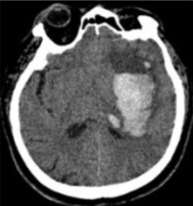
Possible hemorrhagePrioritized
Reading experience takes years to build, and the emergency cases arrive anyway.
A bleed may wait behind several routine scans.
Under pressure, a complete and structured report is hard to write.
In development
Flags suspected intracranial hemorrhage on non-contrast head CT and moves those cases to the top of the reading list.
For: Emergency doctors, on-call radiologists.
In development
Drafts a structured head CT report from a standard template and reminds you of the items that are easy to miss.
For: Radiologists, residents.
Roadmap
Planned after the two tools above.
Target findings in development. Not validated for clinical use.
Next
The doctor always makes the final call.
Reading list
Priority RoutineReview soonUrgent
Draft report
Hyperdense focus in the left basal ganglia. Ventricles and midline: for the doctor to confirm.
Suspected intraparenchymal hemorrhage.
Check: midline shift
Draft. Not yet reviewed by a doctor.
These are design goals for tools that are still in development.
Designed for an on-premise option inside the hospital network, in line with Vietnam's Law on Personal Data Protection.
Designed to work alongside the PACS your hospital already uses.
Drafts follow the report templates your department knows.
Talk to us by Zalo, phone or in person.
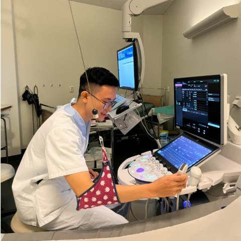
Albert Pham
Founder. Radiology Resident, University of Medicine and Pharmacy at Ho Chi Minh City
I've seen how much a few years of experience change a night shift. ApiVision is my way of giving that to every young doctor.
Interested in joining? We're looking for AI engineers and clinical collaborators.
Leave your details and we will get back to you. You can also write directly to [email protected].